
All Wales Cellular Bioenergetics Analysis Platform
Monitoring mitochondrial health is crucial to drug discovery and development. In particular, drug safety can be monitored and improved through mitochondrial metabolic functional characterisation and help reduce late stage attrition. The primary role of mitochondria is to produce cellular energy in the form of adenosine triphosphate (ATP) via a process called oxidative phosphorylation.
Breakdown in mitochondrial function might be a key factor leading to age-related disease and their malfunction is a prominent finding in a range of human diseases including cancer and neurodegeneration. Evaluation of mitochondrial function is critical to determining underlying metabolic mechanisms involved in human disease and in drug development so has broad impact for improved understanding of multiple diseases and development of the drugs to treat these.
The XFe24 Cellular Flux Analyzer is a high throughput plate based real-time mitochondrial bioenergetic measuring system that can assess oxidative phosphorylation, glycolysis and fatty acid oxidation of any cell type (http://www.seahorsebio.com) (Figure 1). Specifically, the XFe24 Cellular Flux Analyzer measures the oxygen consumption rate (OCR, a specific measure of oxidative phosphorylation and fatty acid oxidation) and the extracellular acidification rate (ECAR, a specific measure of glycolysis) after the sequential injection of mitochondrial stress-inducing or glycolysis regulating reagents (Figure 2). The Seahorse XFe24 Cellular Flux Analyser (Seahorse Bioscience) is a recent technological innovation that enables high efficiency monitoring of mitochondrial cellular function in both intact cells and isolated mitochondria.
Figure 1. (left) The Extracellular Flux Analyser from Seahorse Bioscience.
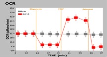
Figure 2. (right) (Seahorse XF data showing the effect of OCR (oxygen consumption rate) in mitochondria after the injection of different drugs to intact test cells. Control cells were sham injected.
For further details please contact Professor Cathy Thornton on (01792) 602122 (c.a.thornton@swansea.ac.uk).